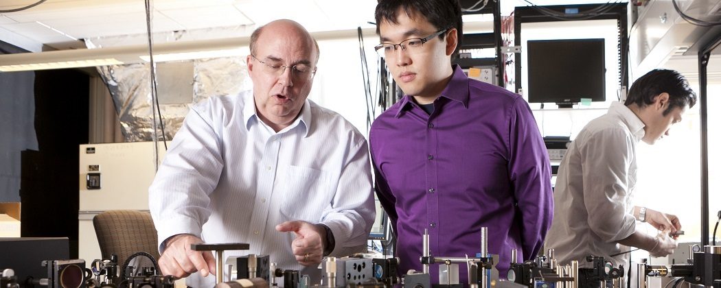
The connection between biological tissue and historical artwork may seem strange at first, but the tools in our lab are excellent for performing three-dimensional imaging of both. The key is depth. Just as the structure of a melanoma changes as you look deeper into human skin, the structure of a painting changes as you look through the visible layer to see what lies beneath. By coupling two different colored pulses with a variable interpulse delay we can take 3-D chemical specific images of historical art pigments.

Jointly funded by NSF Project CHE-1309017, the Center for Molecular and Biomolecular Imaging, and the North Carolina Museum of Art.
Publications
T.E. Villafana, W.P. Brown, J.K. Delaney, M. Palmer, W.S. Warren, and M.C. Fischer, “Femtosecond pump-probe microscopy generates virtual cross-sections in historic artwork”, Proc. Nat. Acad. Sci., (2014) doi: 10.1073/pnas.1317230111
Media Coverage
3D Imaging Reveals How Paintings Were Made | Science
Laser Looks Under the Surface of Art | Nature
Pinpointing Pigments in 3-D | Chemistry & Engineering News
Art Under a New Wavelength | UNC-TV
New Use for Laser in the Art World | phys.org
Uncovering Art | NSF Science 360
Lasers ID Ancient Artists’ Intent | Research @ Duke
